
Home
Common Techniques
Classroom Experiments
Virtual Experiments
Tutorials
Games
Glossary
Links
Publishing
Opportunities
About This Site
Contact Us
ZFIN
Cite Us
Stages of Zebrafish Development
Open protocol in PDF
Download the pre-lab worksheet
Download staging images, or go to the Virtual Staging Experiment
Background Information
The purpose of this laboratory is to properly identify the developmental stages of various zebrafish embryos. Microscopy techniques will be developed and enhanced during this lab as well.
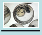
The zebrafish is an important model organism in the study of vertebrate development. This model system is especially useful in forward genetic studies. This means that a researcher starts with a mutant phenotype (for example, a mutant in which the neural tube does not close correctly or some other genetic variable) and then looks for the gene that causes this mutation. Another benefit of studying the embryonic development of zebrafish is that the chorion, or egg shell, is clear; thus, zebrafish development can be seen and observed clearly under a simple microscope. The somites can also be counted if darkfield microscopy is used. Other advantages of this model organism include rapid development, ability to raise a large amount of fish in a small space, and easy transplantation of cells.
As previously mentioned, the embryos develop rapidly; in fact, cell divisions occur approximately every 15 minutes and the first movements begin in under 24 hours. Additionally, after 72 hours of development, the larva begins to seek food on its own and displays active avoidance behaviors.
This next section will describe the stages of zebrafish development beginning with fertilization. The names of the stages will be italicized. When Fertilization occurs, all of the cellular factors (proteins and organelles) needed for early development are already loaded into the egg. The first period of cellular divisions is called the zygote period and it lasts from 0-.75 hours. In this period, the cytoplasm moves toward the animal pole to form the blastodisc, which is where future cell divisions will occur. The blastodisc of the telolecithal egg (meaning that the majority of the egg is yolk, which classifies zebrafish development) then divides into 2 cells at about .75 hours.The next period is called the cleavage period and it lasts until the 64-cell stage; the blastomeres (which are the dividing cells) divide equally about every 15 minutes.
After cleavage is the blastula period. During this period, at about the 10th cleavage, the egg goes through mid-bastula transition (MBT), which is when zygotic gene transcription occurs. Another important event that occurs is the formation of the yolk synctial layer (YSL) which occurs when the cells that are closest to the yolk fuse to it. The last thing that occurs during the blastula period is the beginning of epiboly. This is a type of cell movement in which all embryonic cells move so that the ectoderm, or outer layer, is on the outside of the forming embryo.
At 50% epiboly, the gastrulation period begins. Gastrulation is characterized by its vast amount of cell migrations; the purpose of this migration is to make sure each germ layer (endoderm, mesoderm, ectoderm) is in the right place so that bodily organs and tissues can form in the correct location. For example, the ectoderm will form parts of the body such as the brain and skin. The mesoderm will form parts of the body such as somites and mesenchyme which will eventually form tissues such as muscles. Finally, the endoderm will eventually form into the lining of the digestive tract. As you can see, gastrulation is a very important stage of zebrafish development, and it occurs from about 5.3- 22 hrs.
Following gastrulation, the segmentation period occurs in which the 3-somite to 26-somite stages occur. Somites eventually become dermis, vertebrae and skeletal muscle. One important technique that this classroom experiment will teach you is how to look at an embryo in this segmentation period and count the number of somites to determine the exact stage.
Next, from 24-48 hours, is the pharyngula period. One major occurrence in this period is that the body axis begins to straighten, rather than hugging the yolk sac as it has done previously; additionally, the fins begin to develop. Lastly, the Hatching Period occurs from about 48-72 hours. In this period, primary organ systems develop and cartilage development begins.
Instructor Information
This is a beginner level experiment ideal for students who have a limited background in zebrafish development. This lab can easily be tailored to fit into the development section of an Introductory Biology course, especially because it can be performed in large class sizes. If a limited number of embryos and/or microscopes are available, students may be able to work in groups.
Materials needed
- Wild type zebrafish embryos in various stages
- Dissecting stereomicroscopes
- Compound microscope
- Fine forceps
- Materials for making embryo loops (capillary tubes, fishing line and superglue)
- Glass slides
- MS-222 or Tricaine for anesthetizing the embryos
- Methylcellulose for mounting the embryos.
Techniques Used
- Introduction to dissecting stereomicroscope
- Dechorionation
- Creating embryo loops
- Introduction to Compound Microscope
- Anesthetizing embryos
- Mounting embryos in methylcellulose
- Brightfield vs. Darkfield Microscopy